by
Lauren Dubinsky, Senior Reporter | January 31, 2018
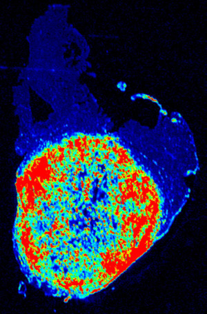
The tumor sharply illuminated
by the fluorescent nanosensor
Researchers at the UT Southwestern Simmons Cancer Center in Dallas, Texas developed a digital nanosensor that illuminates cancer tissue during surgery.
The team is gearing up to test whether this new technology can improve the accuracy of cancer surgeries in order to reduce cancer recurrence and surgical morbidity.
Since tumors are acidic, they secrete acids into the surrounding tissue and that alters the pH level. The nanosensor works by amplifying pH signals in tumor cells to more accurately differentiate them from healthy cells.


Ad Statistics
Times Displayed: 2436
Times Visited: 4 Stay up to date with the latest training to fix, troubleshoot, and maintain your critical care devices. GE HealthCare offers multiple training formats to empower teams and expand knowledge, saving you time and money.
The nanosensor technology and the camera used to view the fluorescent cells are under development by OncoNano Medicine Inc. The current nanosensor is also compatible with existing OR cameras such as the NOVADAQ's SPY Elite system.
The clinical fluorescence imaging camera is much different from other imaging modalities commonly used to evaluate cancer, such as PET and SPECT.
"These [fluorescence cameras] can be used intraoperatively, do not require radioactivity, and are much less expensive," Dr. Baran Sumer, surgeon and professor at the Simmons Cancer Center, told HCB News. "They are also widely available with existing systems in place throughout most hospitals in the U.S."
In December 2016, the team published research in
Nature Biomedical Engineering that demonstrated the nanosensor’s ability to illuminate tumor tissue in multiple mouse models. The next step is a clinical trial.
They plan to initially test it on breast cancer patients at the University of Groningen in the Netherlands. The patients will be injected intravenously with the nanosensor medication 12 hours prior to surgery and the tumors will remain illuminated for one to two days.
This is not the first time that a research team made cells glow — the Washington University School of Medicine developed a
pair of goggles that illuminate cancer cells in 2014 and the University of Pennsylvania is working on an
imaging tool that uses a targeted fluorescent dye to light up benign brain tumors that can cause blindness, hormonal disorders, and in some cases, gigantism.
The team expects to have data on breast cancer and colon cancer surgery by fall this year, and plan to initiate Phase II clinical trials in the U.S. and elsewhere.


