by
John R. Fischer, Senior Reporter | May 28, 2021

Deep-learning algorithm can detect both cancer and risk for cardiovascular disease from the same low-dose CT scan
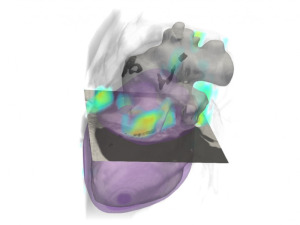
Clinicians at Massachusetts General Hospital and engineers at Rensselaer Polytechnic Institute have developed a deep learning algorithm capable of screening for cardiovascular disease risk and cancer from the same scan.
Applied to low-dose CT, the AI solution is designed to expedite the diagnosis process, accelerate treatment and improve patient outcomes, while eliminating the need for additional scanning and subsequently, more radiation exposure.
“Recent studies have shown that the patients diagnosed with cancer have a much greater risk of CVD mortality than the general population. Nevertheless, when the cancer risk population receives cancer screening, their potential CVD risk may be overlooked. Our work shows that deep learning can convert LDCT for lung cancer screening into a dual-screening quantitative tool for CVD risk estimation for this high-risk group,” Pingkun Yan, an assistant professor of biomedical engineering and member of the Center for Biotechnology and Interdisciplinary Studies (CBIS) at Rensselaer, told HCB News.


Ad Statistics
Times Displayed: 1023
Times Visited: 5 Fast-moving cardiac structures have a big impact on imaging. Fujifilm’s SCENARIA View premium performance CT brings solutions to address motion in Coronary CTA while delivering unique dose saving and workflow increasing benefits.
Developed at Rensselaer and tested at Massachusetts General Hospital, the algorithm proved highly effective in analyzing the risk for cardiovascular disease and related mortality in high-risk patients undergoing low-dose CT, and was equally sufficient as radiologists in analyzing these images. It also nearly mirrored the performance of dedicated cardiac CT scans when applied to an independent data set collected from 335 patients.
The solution was developed, trained and validated on data from more than 30,000 low-dose CT images from a large data set from the National Lung Screening Trial (NLST), which has affirmed that early detection and treatment with low-dose CT can reduce lung cancer mortality. Its design included making sure it could filter out unwanted artifacts and noise and extract features needed for diagnosis. Researchers also validated it using another 2,085 NLST images.
Heart disease and cancer share common risk factors, including tobacco use, diet, blood pressure and obesity. A
multi-institutional study from 2019 found cancer put patients at higher risk of dying from cardiovascular disease based on an analysis of three million cancer cases and 28 different types of malignancies.
Yan and his colleagues are planning more multi-vendor, multi-site studies to further validate the algorithm.
The findings were published in
Natures Communications.
Back to HCB News

