by
John R. Fischer, Senior Reporter | January 20, 2022
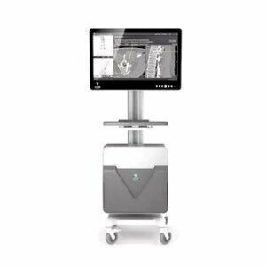
VUZE system (Photo courtesy of VUZE Medical)
VUZE Medical has scored FDA 510(k) clearance for the VUZE system, a software-only solution designed to assist in X-ray guidance during spinal stabilization surgeries, particularly short spinal segment procedures.
Spinal stabilizations correct instabilities or deformations. A third of the total annual number are performed in the U.S. and include vertebral fixation with pedicle screws, vertebral fixation coupled with fusion and vertebral augmentation with synthetic or biological cement. Short-segment procedures make up approximately 80% of stabilizations and are often performed manually and in the OR, where the only imaging required is standard 2D X-ray.
The VUZE system offers preoperative surgical planning, including implant size, entry location and trajectory determination. It also can be used for intraoperative guidance and to confirm that patients are positioned correctly.


Ad Statistics
Times Displayed: 18953
Times Visited: 49 Stay up to date with the latest training to fix, troubleshoot, and maintain your critical care devices. GE HealthCare offers multiple training formats to empower teams and expand knowledge, saving you time and money.
It is meant for use in outpatient and ambulatory settings, according to VUZE Medical’s CEO David Tolkowsky. “Rising cost pressures and a growing aversion toward hospitalization in the age of COVID-19 are accelerating a shift of short-segment surgeries from inpatient to outpatient and from outpatient to ambulatory settings. In such settings, reliance on X-ray guidance is particularly high.”
The solution merges intraoperative X-ray with preoperative CT to create cross-sectional images that surgeons need but lack during procedures. It does this by producing intra-operative 2D images onto axial and sagittal views generated from the patient’s standard preoperative 3D scans.
The hardware is an on-cart, off-the-shelf PC. It has no sensors, cameras or reference arrays and requires no calibrations or lines of site. It also can be used with standard surgical tools and implants without tool add-ons or modifications.
Drs. Ory Keyman and Elias Haddad at Rambam Medical Center in Israel were the first to use this system, and treated a number of their patients with it successfully.
The company expects it to be used for the first time on patients in the U.S. in mid-2022.


