by
John R. Fischer, Senior Reporter | April 15, 2022

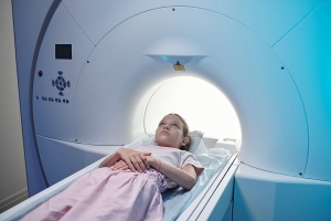
A new deep-learning reconstruction technique can reduce dose used in pediatric CT scans, while still maintaining or improving image quality.
Using deep learning-based reconstruction, Japanese researchers have reduced the dosage in pediatric CT exams while still maintaining the same or even improving on the image quality of those using interactive reconstruction algorithms.
Because children are more sensitive to ionizing radiation, clinicians use the lowest possible dose when scanning them. One way to do that is to decrease tube voltage with iterative reconstruction. But lowering voltage increases image noise and impairs detection of low-contrast objects, especially when using reduced slice thickness to assess a child’s small anatomic structures.
Hybrid iterative reconstruction (HIR) and model-based iterative reconstruction (MBIR) can reduce noise and artifacts but do not preserve noise texture, low-contrast spatial resolution and low-contrast object detectability as well when decreasing dose.


Ad Statistics
Times Displayed: 2648
Times Visited: 16 Fast-moving cardiac structures have a big impact on imaging. Fujifilm’s SCENARIA View premium performance CT brings solutions to address motion in Coronary CTA while delivering unique dose saving and workflow increasing benefits.
An emerging technique for CT image reconstruction, DLR uses a convolutional neural network to generate low-noise, high-quality images in short time frames,
reports Physicsworld. The team applied the application to low-tube-voltage exams and reduced noise without degrading the noise texture and image sharpness. Compared to HIR and MBIR, DLR decreased radiation dose by approximately 50% and still preserved or even improved image quality and task-based object detectability of the scans. “The findings may be applied to achieve substantial radiation dose reductions in pediatric CT in comparison with current IR techniques,” wrote the researchers in their study.
The team tested and compared all three applications through a retrospective analysis of scans for 65 children, ages six and under. All underwent contrast-enhanced abdominal CT, with 31 assessed with a standard protocol and 34 with a lower-dose protocol. Tube voltage for all exams was 80 kVp (unlike the standard 120 kVp used in adults), and the AiCE (Advanced intelligent Clear-IQ Engine) Body-sharp algorithm was applied for DLR.
They found that the size-specific dose estimate (SSDE) for the lower-dose protocol group was 54% lower than that of the standard group (1.9±0.4 versus 4.0±1.0 mGy). They also reported significantly higher subjective scores for noise magnitude, noise texture, edge sharpness and overall quality than standard HIR, low-dose HIR and low-dose MBIR.
DLR also outperformed both iterative reconstruction methods in a phantom analysis used to assess image quality at a wider range of dose settings with a 20 cm cylindrical phantom. The same CT scanner and image parameters for the clinical patients were used here, with eight fixed tube currents applied to create doses relevant to clinical pediatric CT.
The findings were published in the
American Journal of Roentgenology.

