Breast PET/CT scanner
will be able to improve
breast cancer imaging
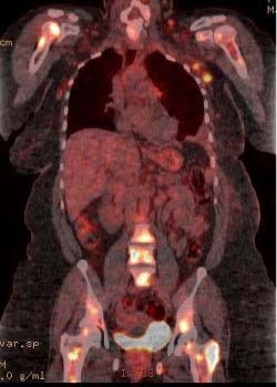
NEW ORLEANS, LA - The first patient scans from a custom-built scanner combining positron emission tomography (PET) and computed tomography (CT) technologies indicate that these scans could significantly improve breast cancer imaging capabilities and lead to more targeted treatment options, according to researchers at SNM's 55th Annual Meeting. The prototype scanner is designed to help physicians determine stages of breast cancer in patients already diagnosed with the disease, rather than as a mammography screening tool.
"The use of dedicated breast PET/CT scanners could really open up new possibilities in treatment for women with breast cancer," said Ramsey Badawi, assistant professor of radiology, University of California-Davis Medical Center, Sacramento, and investigator for the study, First Human Images from a Dedicated Breast PET/CT Scanner. "Using this noninvasive technology, physicians can get much more accurate images of tumors-especially small tumors-than conventional full-body PET scans. This will enable physicians to determine the stage of the cancer and determine courses of treatment more accurately."
In addition, the technology could eventually be used to detect early whether drug treatments are effective in individual patients. "One great advantage for doctors will be to better plan breast cancer surgeries," said Badawi. "Using a PET/CT scan, doctors should be able to determine whether drug treatments are working before performing surgery, thus eliminating unnecessary mastectomies. Medical professionals will also have a much better sense of the exact location and size of tumors."


Ad Statistics
Times Displayed: 45539
Times Visited: 1299 Ampronix, a Top Master Distributor for Sony Medical, provides Sales, Service & Exchanges for Sony Surgical Displays, Printers, & More. Rely on Us for Expert Support Tailored to Your Needs. Email info@ampronix.com or Call 949-273-8000 for Premier Pricing.
The technology also can be used to measure the effectiveness of new drugs and molecular imaging agents for detecting and treating breast cancer, another important component to advancing individualized medicine for patients. In addition, the dedicated breast PET/CT scanner can detect the difference between malignancies and benign tissues such as cysts or scars, eliminating many unnecessary biopsies.
The prototype breast PET/CT scanner studied by Badawi and his team consists of two adjustable planar heads. Patients lie prone as the PET and CT systems rotate around the freely suspended breast. PET and CD data are used to produce detailed three-dimensional images. Many women could also find the new technology more comfortable, because it does not require the breast to be compressed during the scan.
The first patient to be scanned with the dedicated breast PET/CT scanner was a 49-year-old woman with a palpable mass on her right breast. The dedicated breast PET/CT scan accurately detected breast cancer, which was later confirmed by biopsy. The results indicate that PET and CT images of the uncompressed breast are accurate and provide clinically relevant data.
Scientific Paper 97: S.L. Bowen, A. J. Chaudhari, Y. Wu, L. Fu, J. Qi, S.R. Cherry, Biomedical Engineering; N. Packard, G Burkett, J.M. Boone, R.D. Badawi, Radiology, University of California-Davis, Sacramento: "First Human Images From a Dedicated Breast PET/CT Scanner."
About SNM-Advancing Molecular Imaging and Therapy
SNM is an international scientific and medical organization dedicated to raising public awareness about what molecular imaging is and how it can help provide patients with the best healthcare possible. SNM members specialize in molecular imaging, a vital element of today's medical practice that adds an additional dimension to diagnosis, changing the way common and devastating diseases are understood and treated.
SNM's more than 16,000 members set the standard for molecular imaging and nuclear medicine practice by creating guidelines, sharing information through journals and meetings, and leading advocacy on key issues that affect molecular imaging and therapy research and practice. For more information, visit www.snm.org.


