Conventional MR scanners require around 1,500 liters of liquid helium to function — but with the introduction of deep learning software, that dependency on a nonrenewable resource is rapidly diminishing.
In recent years, major imaging OEMs like
Philips and
Siemens have introduced scanners that run on one liter of helium or less,
Hyperfine has commercialized a head-only MR that requires no helium... and now researchers in China are touting 3T-quality whole-body MR images obtained without any helium.
The Hong Kong team has published findings using a novel ultralow field (ULF) whole-body MR scanner that combines deep learning with a permanent and compact 0.05-Tesla magnet to dramatically reduce operational costs, equipment footprint, and barriers to access for patients and providers.


Ad Statistics
Times Displayed: 365537
Times Visited: 21119 MIT labs, experts in Multi-Vendor component level repair of: MRI Coils, RF amplifiers, Gradient Amplifiers Contrast Media Injectors. System repairs, sub-assembly repairs, component level repairs, refurbish/calibrate. info@mitlabsusa.com/+1 (305) 470-8013
Traditional high-field (1.5T, 3T, and in some cases 7T) MR scanners — which run on powerful magnets and superconducting liquid helium, and require extensive shielding and power consumption — are costly and complicated machines, putting them out of reach for many rural medical offices, as well as low- and middle-income countries.
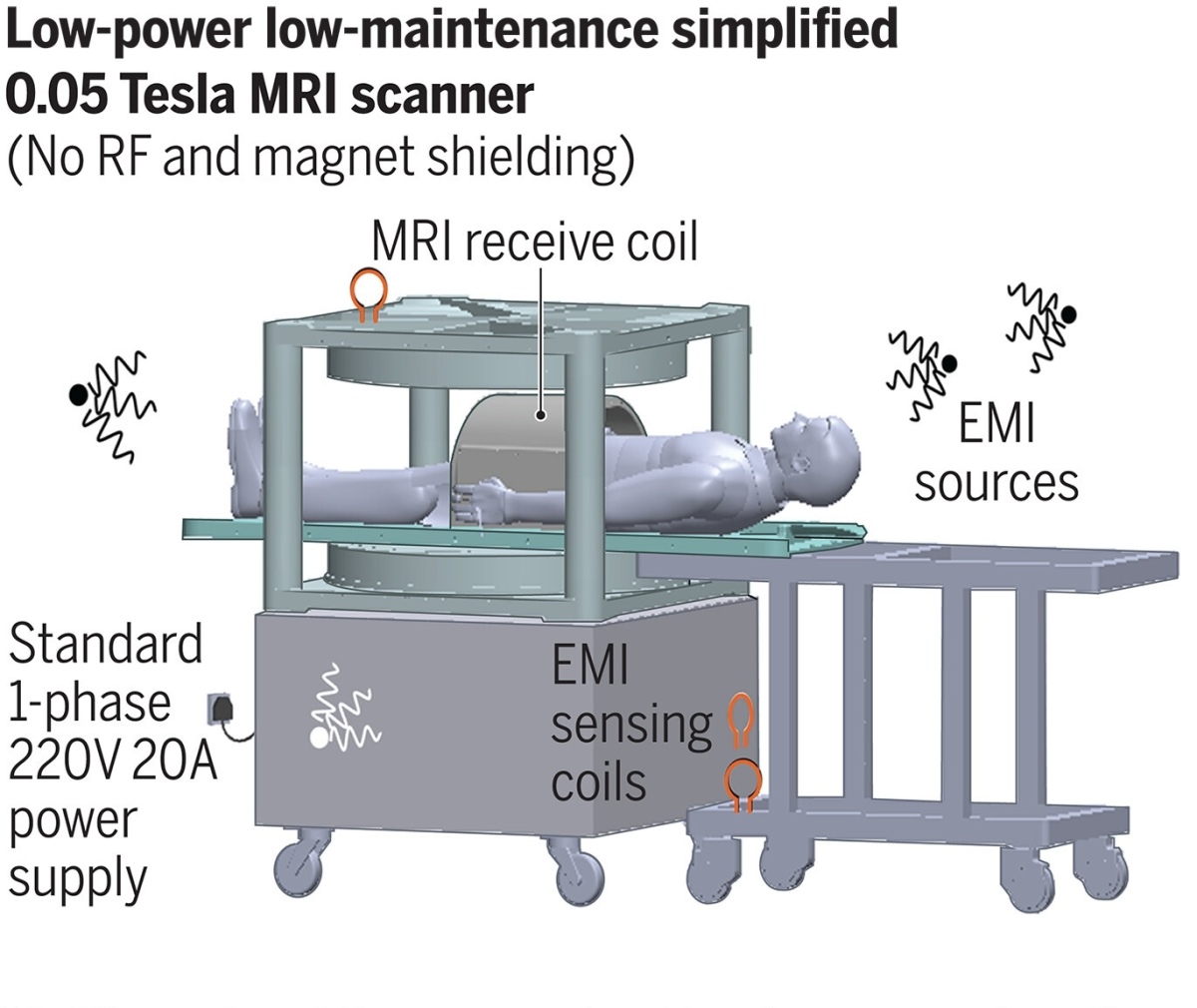
The ULF MR system, by contrast, is compact, requires no shielding, no helium, and operates on a standard wall power outlet. It uses only 1,800 watts of power during scanning, (and 300 watts idle) whereas conventional MR scanners often consume 25,000 or more.
Prototype of a low-cost, low-power, compact, and shielding-free imaging system using an open 0.05 Tesla permanent magnet. It incorporates active sensing and deep learning to address EMI signals.
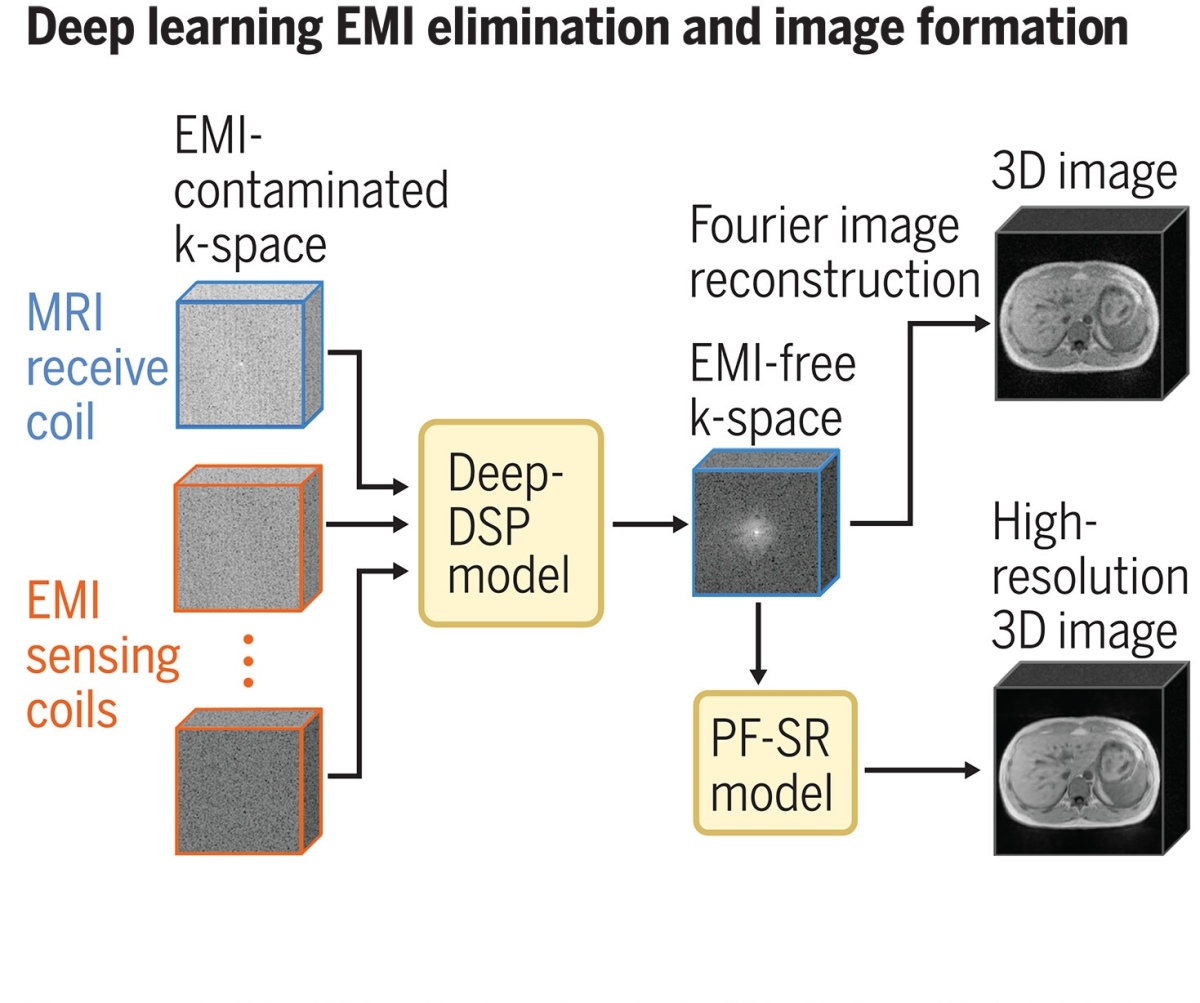
The core innovation of the MR scanner lies in its use of active sensing coils and deep learning algorithms to eliminate electromagnetic interference (EMI), a common challenge with lower field strengths. Clinical protocols tested include various types of imaging, such as T1-weighted, T2-weighted, and diffusion-weighted imaging, each optimized for different anatomical structures with scan times under eight minutes.
"The past technical development of MR relied too much on the hardware and conventional mathematics, but now is an era of computing and large-scale data, and it has been happening in many applied engineering fields," Ed X. Wu, a researcher from the University of Hong Kong who works on the scanner, told HCB News. "Both hardware and smart computing are needed. Our long-term research will help to determine the minimum hardware requirement for clinically useful scanners, especially under point-of-care conditions."
In a
study published in Science, the researchers conducted imaging on healthy volunteers — capturing brain, spine, abdomen, lung, musculoskeletal, and cardiac images — and found they were able to produce clear and detailed imaging on par with that obtained by 3T systems currently used in the clinic:
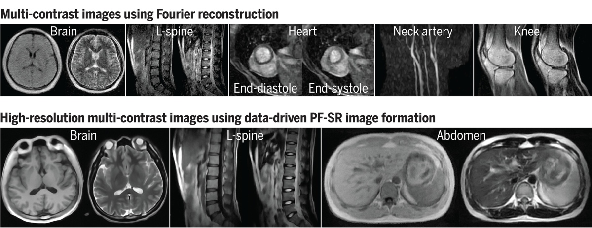
The brain images showed various brain tissues whereas the spine images revealed intervertebral disks, spinal cord, and cerebrospinal fluid. Abdominal images displayed major structures like the liver, kidneys, and spleen. Lung images showed pulmonary vessels and parenchyma. Knee images identified knee structures such as cartilage and meniscus. Cardiac cine images depicted the left ventricle contraction and neck angiography revealed carotid arteries.
The deep learning model employed in the ULF MR scanner utilizes a data-driven image formation method, which integrates image reconstruction and three-dimensional multiscale super-resolution to leverage the homogeneous human anatomy and image contrasts available in large-scale, high-field MR data. As the technology matures, it could pave the way for widespread deployment in hospitals and clinics, substantially reducing the logistical and financial challenges associated with traditional MR systems.
In a
related commentary published in the same issue of
Science, Udunna Anazodo and Stefan du Plessis noted sobering limitations and challenges that must be addressed before low-field MR can be widely applied for clinical use, but also acknowledged the tremendous promise it holds.
"This machine costs a fraction of current clinical scanners, is safer, and needs no costly infrastructure to run," they wrote. "Although low-field machines are not capable of yielding images that are as detailed as those from high-field clinical machines, the relatively low manufacturing and operational costs offer a potential revolution in MR technology as a point-of-care screening tool."
(Top) Typical images of various anatomical structures using conventional image reconstruction. (Bottom) High-resolution images using deep learning image formation by harnessing large-scale high-field MR data.
The ULF MR uses two permanent magnet plates, above and below the body, in an open configuration, which Anazodo and du Plessis described as "bulky," adding that the scanner "acquires coarser images at longer scan times compared with other portable low-field options."
While development is ongoing, Wu is confident that the research he and his colleagues are doing will help usher in a new era where MR imaging is more accessible to everyone.
"I strongly believe that low-field MR can be and should be used like ultrasound in healthcare, in both developed and developing countries," Wu said. "I think some visionary investors, together with new generation of scientists, will eventually make this happen."


 (1)
(1)


jon naude
The 0.05T MRI images
May 21, 2024 01:31
As a radiologist and oncologist I'm very impressed with the imaging from the low-energy MRI. This will bring the costs down dramatically and could literally put an MRI in every practice. I'm sure the established MRI, CT and ultrasound centres will fight it, but MRI is one of the modalities that are pushing up the price of health care because more and more drs and patients are demanding the better imaging. For breast MRI we still have to send our breast cancer patients to a centre hundreds of miles away.
to rate and post a comment
Jason Taylor
re: The 0.05T MRI images
May 23, 2024 10:57
" I'm sure the established MRI, CT and ultrasound centres will fight it,"
The authors will be given tedious teaching assignments and funding cuts. The tech was very predictable. But no, patients will have to get their dose of ionizing radiation, the CT scan "gold standard" war path must continue.
to rate and post a comment