by
Barbara Kram, Editor | September 11, 2006
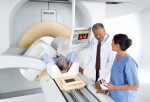
Philips' Precedence 64-slice
SPECT/CT
Andover, Mass. - Philips Medical Systems, a division of Royal Philips Electronics (NYSE: PHG, AEX: PHI), is showcased advances in its portfolio of leading nuclear cardiology solutions at the recent American Society of Nuclear Cardiology (ASNC) annual meeting (September 7-9 in Montreal, Canada). The Precedence 64-slice SPECT/CT is the only hybrid-imaging system that delivers comprehensive cardiac management on a single imaging platform, and the latest version of Xcelera extends its patient-centric approach to image and information management to the nuclear cardiology domain. Philips also displayed the GEMINI TF PET/CT system to feature advances in evaluation and analysis of cardiac disease.
Other highlights include the 1,000th installation of the CardioMD gamma camera and data validating the benefits of computed tomography (CT) angiography.
Hybrid-imaging capabilities of Precedence 64 reinforce Philips leadership in SPECT/CT


Ad Statistics
Times Displayed: 45539
Times Visited: 1299 Ampronix, a Top Master Distributor for Sony Medical, provides Sales, Service & Exchanges for Sony Surgical Displays, Printers, & More. Rely on Us for Expert Support Tailored to Your Needs. Email info@ampronix.com or Call 949-273-8000 for Premier Pricing.
The Precedence family of hybrid-imaging SPECT/CT is one of the industry's most advanced imaging systems capable of dedicated SPECT and diagnostic CT as well as sophisticated hybrid-imaging. The introduction of the 64-slice Precedence advances the capability of the platform by performing uncompromised cardiac imaging on patients who can benefit from the imaging procedure.
"CT improves SPECT imaging on multiple levels," said John Mahmarian, M.D., director of nuclear cardiology at The Methodist Hospital in Houston, Texas. "For patients with normal myocardial perfusion scans, equivocal nuclear studies or abnormal myocardial perfusion scans, hybrid SPECT/CT can improve care tremendously by helping identify early disease states and clarify diagnostic ambiguity."
The Precedence 64-slice SPECT/CT can produce CT-based attenuation correction and perform advanced cardiac CT procedures such as calcium scoring and CTA in one episode of care, on one system. It can also produce SPECT myocardial perfusion imaging in half the time of conventional scanners.
Xcelera expands into nuclear cardiology domain with new modalities, integrated workflow and patient-centric work-space design
Philips is introducing a new release of its integrated cardiovascular information solution, the Philips Xcelera R2.1, which now integrates exam results from all key cardiology subspecialties - interventional cardiology, cardiovascular ultrasound, ECG, nuclear cardiology, cardiac CT, cardiac MR and electrophysiology. This advanced cardiovascular solution for documentation, viewing, quantification and reporting tasks, provides clinicians with access to relevant images and information on patients across the hospital from a single workspace.


