by
Lauren Dubinsky, Senior Reporter | March 04, 2016
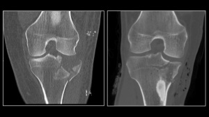
An ultra-low dose CT
scan of a fracture
of the tibial plateau (left)
compared to a conventional
dose CT scan.
Courtesy of NYU Langone
Medical Center
When it comes to CT imaging, radiation is still a major concern among physicians, but new research out of NYU Langone Medical Center may put an end to that. Researchers at the medical center developed a CT protocol for evaluating joint fractures that generates one-fourteenth the amount of normal CT radiation without having an effect on image quality.
“We found we were able to obtain nearly the exact same types of images for various fractures and locations without sacrificing any diagnostic accuracy in most cases,” Dr. Kenneth A. Egol, senior author of the paper and professor of orthopaedic surgery at NYU Langone Medical Center, told HCB News.
In the past, Egol and his colleagues found that they could identify joint injuries with the use of CT. They then conducted a study demonstrating that CT could identify a traumatic arthrotomy of a joint better than a saline load, which used to be the standard.


Ad Statistics
Times Displayed: 346433
Times Visited: 21062 MIT labs, experts in Multi-Vendor component level repair of: MRI Coils, RF amplifiers, Gradient Amplifiers Contrast Media Injectors. System repairs, sub-assembly repairs, component level repairs, refurbish/calibrate. info@mitlabsusa.com/+1 (305) 470-8013
“When we presented that work, one of the criticisms we received was that we were subjecting patients to a high dose of radiation for this diagnostic test,” said Egol. “Our rationale at the time was that the saline load test was a painful, invasive procedure using a needle, and that we would trade a bit of radiation for lack of invasive procedure.”
But as they thought more about it, they realized that they might be able to decrease the amount of radiation in the CT while also maintaining the same diagnostic accuracy. They worked with their department of radiology and found that they could in fact obtain good-quality images and reduce the radiation dose.
They then asked themselves — if we can get a CT scan that shows us good bony detail and is safer, then why shouldn't we be doing it on everybody, not just these arthrotomy cases? That led them to develop the Reduced Effective Dose Using Computed Tomography in Orthopaedic Injury (REDUCTION) protocol.
From August 2014 to March 2015, 50 patients presenting clinical symptoms of joint fractures underwent ultra-low dose radiation CT scans. The researchers compared those scans to a sample of age-matched, similar fracture injuries in which the patients were evaluated with standard CT.
They found that with ultra-low dose CT they could achieve 98 percent sensitivity and 89 percent specificity. They also found that those findings were comparable to standard CT scans and the image quality was rated by orthopaedic surgeons as moderate to near perfect.
Egol thinks that the protocol would be ideal for pediatric fracture evaluation if the fracture is known. However, it may not be as useful in detecting occult fractures, evaluating tumors and other lesions and for applications such as those that might be used in spinal surgery.
In order for the protocol to be implemented on a widespread level outside of NYU Langone Medical Center, it’s going to take education and wide publicity among practicing orthopaedic physicians, emergency room physicians, radiologists, pediatricians and anyone else who would order a CT scan.
Going forward, the researchers will continue to evaluate the patients who underwent ultra-low dose CT scans to ensure that patient outcomes haven’t changed. They hope to eventually expand the protocol to other departments as well.


