by
John W. Mitchell, Senior Correspondent | September 19, 2016
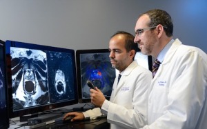
Dr. Daniel Costa, left, and Dr. Ivan
Pedrosa, right, review multiparametric
MRI images
It took three years, but a research team at UT Southwestern Medical Center in Dallas has expanded its initial research on 400 patients and has performed its 1,000th MRI-Transrectal Ultrasound (MRI-TRUS) 3-D fusion procedure this month.
The researchers found that the procedure improves the detection of clinically significant prostate cancer.
"Our previous study with approximately 400 men has shown that, compared with systematic (conventional, 12-core) biopsy, targeted MRI-TRUS biopsy diagnosed 11 percent more intermediate-to-high risk and 16 percent fewer low-risk tumors," Dr. Daniel Costa, assistant professor of radiology at UT Southwestern Medical Center, told HCB News.


Ad Statistics
Times Displayed: 346433
Times Visited: 21062 MIT labs, experts in Multi-Vendor component level repair of: MRI Coils, RF amplifiers, Gradient Amplifiers Contrast Media Injectors. System repairs, sub-assembly repairs, component level repairs, refurbish/calibrate. info@mitlabsusa.com/+1 (305) 470-8013
He said that moving forward, the research team — also composed of physicians from the urology and pathology departments at UT — will need to better evaluate the method in other patient populations. This includes determining MRI-TRUS diagnostic performance on the basis of outcome factors, such as disease-free survival and cost-effectiveness.
The researchers have partnered with colleagues in Brazil to conduct two follow-up studies.
According to Costa, their recent expanded study helped confirm several key findings.
"Our work also highlighted its (MRI-TRUS) robustness and consistency," he said. "With these results replicated when using different imaging protocols — such as studies done with and without endo-rectal coil — different image fusion devices, as well as various radiologists and urologists with different levels of expertise."
He added that the use of the MRI-TRUS fusion biopsy has demonstrated that it is a good option for men with high clinical suspicions for prostate cancer, which include elevated and rising PSA blood levels and abnormal digital rectal exams.
Such patients often test negative using conventional TRUS only biopsy. These patients may or may not have prostate cancer and they may or may not have an aggressive form of the disease.
The fused MRI-TRUS image creates a 3-D model, and flags anomalies that could be areas of concern. That helps guide urologists to select biopsy samples to determine whether cancer is present and how severe it may be.
Prostate cancer is the second most common cancer diagnosed in men, after skin cancer. Prostate cancer risk increases with age, with most cases occurring after age 60. According to the National Cancer Institute (NCI), about 180,890 men will be diagnosed this year, and about 14 percent of men will be diagnosed sometime during their lifetime.
"Patients diagnosed at a later stage of disease, or with a more aggressive cancer, have lower rates of survival, making it vital that we quickly identify those who are at the highest risk," said Claus Roehrborn, Chair of the UT Urology Department and a member of the MRI-TRUS study team.
Another team at Virginia Commonwealth University Medical Center is
also using a MRI-TRUS method to obtain random tissue samples from 12 cross sections of the prostate to diagnose prostate cancer.


