by
Lisa Chamoff, Contributing Reporter | July 24, 2017

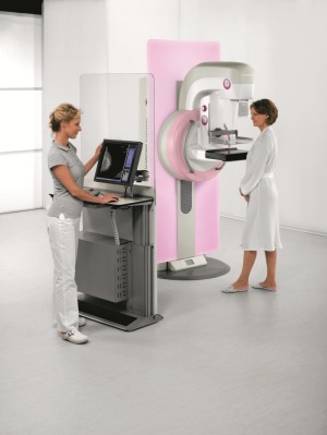
Siemens Healthineers Inspiration with HD Tomo
From the July 2017 issue of HealthCare Business News magazine
While digital 2-D mammography remains a necessary part of cancer screening for the vast majority of women, new technologies are emerging that may help clear the path for digital breast tomosynthesis, also known as 3-D mammography, to become the new gold standard.
Most facilities that screen with tomosynthesis also capture an image via 2-D mammography, in order to give radiologists a look at the entire breast, as well as the individual layers of the breast that tomosynthesis provides.
A few facilities have been studying a new technique that uses the data obtained during tomosynthesis imaging and compresses it to create a synthesized 2-D-like composite image. This, in turn, lowers the radiation dose, as a woman does not need a mammogram in addition to tomosynthesis, which exposes patients to more radiation than digital mammography.


Ad Statistics
Times Displayed: 78287
Times Visited: 2781 Ampronix, a Top Master Distributor for Sony Medical, provides Sales, Service & Exchanges for Sony Surgical Displays, Printers, & More. Rely on Us for Expert Support Tailored to Your Needs. Email info@ampronix.com or Call 949-273-8000 for Premier Pricing.
In a study published last year in the journal Radiology, radiologists at the University of Pennsylvania compared recall rates, biopsy rates, cancer detection rates and radiation dose for more than 15,000 women screened with digital mammography and digital breast tomosynthesis between October 2011 and February 2013, as well as more than 5,000 women screened with digital breast tomosynthesis and synthesized 2-D mammography from January to June 2015.
The study found no significant difference in cancer detection rate for synthesized 2-D mammography and digital breast tomosynthesis versus digital mammography and tomosynthesis, while the average radiation dose was nearly 40 percent lower when the synthetic image was used.
The biopsy rate was also lower when synthesized 2-D mammography was used — 1.3 percent versus 2 percent for the digital mammography and tomosynthesis combination.
“We’ve shown it’s adequate,” says Emily Conant, chief of the division of breast imaging at the Hospital of the University of Pennsylvania and an author of the study. “It further reduces false positives and kept the cancer detection rate the same. If we can create a lower-dose, better mammogram, that’s better.”
Conant says that the 2-D picture is important for looking at a woman’s overall breast density and that some suspicious findings, such as calcium deposits, are more easily seen on a 2-D image. And while some radiologists don’t think the appearance of the synthetic image is as good as a standard 2-D mammogram image, Conant believes the synthesized 2-D mammography and tomosynthesis combination is the future of breast cancer screening.
“In the six years since we’ve implemented tomosynthesis in our clinics, we’ve really seen the impact that it makes,” Conant says. “This whole concept of synthetic imaging is the new kid on the block and it continues to improve. It’s evolving so rapidly. It can only get better.”

