by
Lauren Dubinsky, Senior Reporter | July 26, 2017
Might also help develop new guidelines
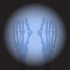
A research team in South Korea found that X-ray imaging can reliably predict bunion recurrence immediately after surgery.
When bunions become severe, painful or interfere with walking, surgery can realign the bone. But studies have reported bunion recurrence rates of up to 25 percent.
Dr. Chul Hyun Park of Yeungnam University Medical Center and Dr. Woo-Chun Lee of Injie University Seoul Paik Hospital set out to determine if X-ray imaging could make a difference.


Ad Statistics
Times Displayed: 316
Times Visited: 2 Keep biomedical devices ready to go, so care teams can be ready to care for patients. GE HealthCare’s ReadySee™ helps overcome frustrations due to lack of network and device visibility, manual troubleshooting, and downtime.
The study included 93 patients undergoing bunion surgery on 117 feet. At an average follow-up of two years, the bunion recurrence rate was 17 percent.
Recurrence was defined as a hallux valgus angle (formed by the toe bone and first metatarsal bone) of 20 degrees or more. Patients with larger preoperative and postoperative HVAs were at higher risk of recurrence.
The researchers found that bunions were 28 times more likely to recur when the postoperative HVA was eight degrees or larger than when the HVA was less than eight degrees.
Other factors related to an increased risk of recurrence included severe bunions with a preoperative HVA of 40 degrees or larger and the position of small bones called sesamoids under the joint shown on postoperative X-rays.
Park and Lee believe that if additional research confirms these results, X-rays taken during surgery may help develop guidelines for the "satisfactory correction" of bunions.


