by
John R. Fischer, Senior Reporter | September 26, 2019
The FDA has given a thumbs-up to the first modules of Siemens’ AI-Rad Companion Chest CT, the intelligent assistant of the AI-Rad Companion platform.
“What we’re trying to do with AI-Rad companion is bring artificial intelligence into routine imaging to help the radiologist,” Peter Shen, VP of Business Development for the Digital Services business at Siemens Healthineers North America, told HCB News. “We’re giving the radiologist an automation tool to process and prepare large amounts of clinical data for interpretation and reduce the growing burden of rising exam volumes.”
The platform is part of the German healthcare giant’s long-term investment to leverage AI in medical imaging. The approved modules of AI-Rad Companion Chest CT will aid clinicians in their interpretations of the thorax, with desired accuracy and precision to optimize clinical operations.


Ad Statistics
Times Displayed: 157
Times Visited: 2 Fast-moving cardiac structures have a big impact on imaging. Fujifilm’s SCENARIA View premium performance CT brings solutions to address motion in Coronary CTA while delivering unique dose saving and workflow increasing benefits.
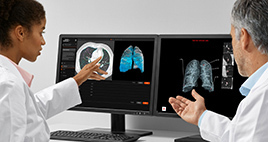
Trained on extensive data sets and annotated by qualified clinical specialists, the algorithms in AI-RAD Companion Chest CT are designed to assess and extract additional clinically relevant information from CT chest images, in addition to that of the primary indication. Among its capabilities are segmentation, measurement and highlighting of key anatomical structures, all of which support quantitative and qualitative analysis.
More specific capabilities include automated detection of lesions, localization of abnormalities, and measurement of lung lesions; quantification of per-lobe, low-attenuation parenchyma; enhanced visualization of lung lesions; automated segmentation of lung lobes and enhanced visualization of low-attenuation parenchyma; segmentation and measurement of maximum diameters of the thoracic aorta; quantification of the total calcium volume in the coronary arteries; and detection of nine anatomical landmarks as identified by American Heart Association (AHA) guidelines.
The assistant can differentiate among diverse structures in the region of the chest, including the lungs, heart and aorta. In addition, the cloud-based solution automatically generates standardized, reproducible, and quantitative reports in Digital Imaging and Communications in Medicine (DICOM) SC format, thereby reducing time spent on manual documentation of findings. Radiologists can access these reports in their PACS systems.
“This initial release of modules is focused on pulmonary and cardiovascular work related to Chest CT," said Shen. "We are in the process of submitting additional modules across other modalities and organs to support further clinical interpretations.”
AI-Rad Companion Chest CT integrates seamlessly within existing clinical workflows. It is also multi-modality and vendor-agnostic, having been tested and validated for CT scanners from Siemens Healthineers, GE Healthcare, and Philips Healthcare.
Forthcoming modules will be showcased at RSNA.

