by
John R. Fischer, Senior Reporter | September 23, 2022
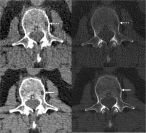
Photon-counting CT and AI for reducing noise are better together at detecting bone disease linked to multiple myeloma, at lower doses, than conventional CT.
Photon-counting CT integrated with AI-based noise reduction capabilities may be more accurate in detecting bone disease associated with multiple myeloma than conventional CT, at lower radiation doses.
Photon-counting CT decreases pixel size and improves spatial resolution in images by converting individual X-ray photons into an electrical signal, and has higher dose efficiency, allowing for ultrahigh-resolution images of large areas of the body to be captured.
Researchers at Mayo Clinic in Rochester, Minnesota, used it to perform whole-body, low-dose scans on 27 patients with multiple myeloma, a white blood cell disease found in bone marrow, and compared it to conventional CT. Approximately 80% of multiple myeloma cases contain areas of bone destruction known as lytic lesions.


Ad Statistics
Times Displayed: 364749
Times Visited: 21098 MIT labs, experts in Multi-Vendor component level repair of: MRI Coils, RF amplifiers, Gradient Amplifiers Contrast Media Injectors. System repairs, sub-assembly repairs, component level repairs, refurbish/calibrate. info@mitlabsusa.com/+1 (305) 470-8013
They also applied a deep learning AI technique developed at Mayo Clinic’s CT Clinical Innovation Center for reducing noise in very sharp photon-counting images. The combination of both technologies improved visualization and showed clinicians more lesions.
“We were excited to see that not only were we able to detect these features of multiple myeloma disease activity more clearly on the photon-counting scanner with deep learning denoising techniques that allowed us to generate thinner image slices, but we were able to detect more lesions than on the standard CT,” said study lead author Dr. Francis Baffour, diagnostic radiologist at the Mayo, in a statement.
Baffour and his colleagues are investigating how they can continue to lower dose and still obtain quality diagnostic CT images with the technology, especially for pediatrics, pregnancy and screenings.
They also are looking into follow-up studies on patients with multiple myeloma precursor status to determine if photon-counting CT can identify bone lesions that would upstage them to active multiple myeloma.
“Our excitement as scientists and radiologists in these results stems from our realization that this scanner could make a difference in the staging of disease, potentially impact therapy choice, and ultimately, patient outcomes,” said Baffour.
The findings were published in
Radiology.


