by
Gus Iversen, Editor in Chief | September 10, 2018
From the September 2018 issue of HealthCare Business News magazine
HCB News: What kind of research did you start off doing with the system prior to scanning the first human patient?
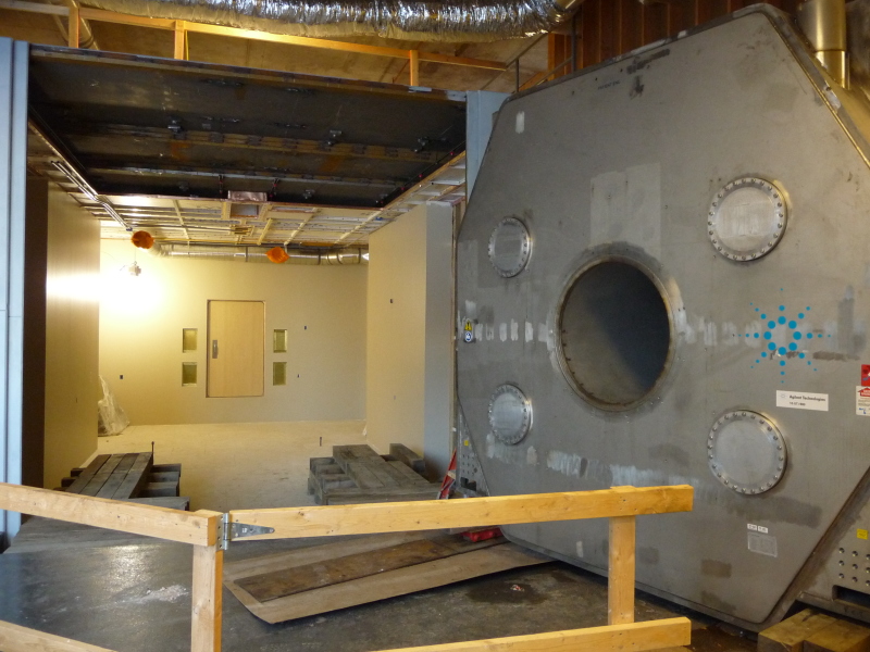
Extensive preclinical imaging was performed to investigate the vestibular, cognitive and physiologic effects of exposure to ultrahigh magnetic fields. The data supported an Investigational Device Exemption (IDE) application submitted to the FDA in order to justify an initial safety study to be performed on this one-of-a-kind system.


Ad Statistics
Times Displayed: 1227
Times Visited: 6 Fast-moving cardiac structures have a big impact on imaging. Fujifilm’s SCENARIA View premium performance CT brings solutions to address motion in Coronary CTA while delivering unique dose saving and workflow increasing benefits.
The IDE and subsequent safety study were required as magnets with field strengths greater than 8 Tesla are no longer considered non-significant risk per FDA guidelines, “Criteria for Significant Risk Investigations of Magnetic Resonance Diagnostic Devices”. Only after the safety study is completed and results are positively reviewed by the FDA will broader clearance be given for more specific studies to investigate the system’s capabilities and pursue answers to biomedical questions.
HCB News: How was the first human patient chosen?
A greater signal-to-noise (SNR) ratio is a key benefit high
field strength magnets may have over conventional MR systems.
The first human imaging studies were performed on healthy controls that passed the strict screening procedure of the ongoing FDA and local IRB (Institutional Review Board) approved safety study. In this initial cohort of subjects, physiological parameters need to be within normal ranges and a health questionnaire is administered and reviewed by a physician to rule out individuals with pre-existing conditions that meet specific exclusion criteria. In addition, subjects need to be free of any metallic implants while meeting both age and weight requirements.
HCB News: What were the first imaging studies performed and how would you describe the image quality compared to a conventional 3 T MR scan?
To date, the studies that have been performed have been in the human torso. This general anatomical region has been the focus, due to the ability to more rapidly validate radiofrequency (RF) coils, which are heavily loaded by the body. Our body array coils consist of elements that both transmit and receive RF energy from the body (i.e., transceivers). Each element is pressed against the body, facilitated by a flexible housing. For head imaging, a similar flexible housing is not feasible, therefore the elements can have varying distance from the head, making the validation of head RF coils more time-consuming.