by
Loren Bonner, DOTmed News Online Editor | September 28, 2012
From the September 2012 issue of HealthCare Business News magazine
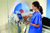
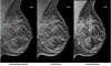
“It’s a slower pace, but there’s an increase in MR breast imaging, abdominal and other body procedures that should soon increase at a faster pace than neuro or musculoskeletal,” says Narayan.
Siemens’ Dhamankar says he sees cardiac MRI growing “because more publications are coming out from the European registry where they talk of its value.”


Ad Statistics
Times Displayed: 109945
Times Visited: 6642 MIT labs, experts in Multi-Vendor component level repair of: MRI Coils, RF amplifiers, Gradient Amplifiers Contrast Media Injectors. System repairs, sub-assembly repairs, component level repairs, refurbish/calibrate. info@mitlabsusa.com/+1 (305) 470-8013
He also sees interventional MRI on the rise, specifically, radiation therapy planning.
“MRI is increasingly being used to better differentiate normal tissue from abnormal tissue, as MRI has excellent soft tissue resolution. For the radiation physicist, to plan dose contours, MRI helps to significantly avoid nerves and vessels, so that complications can be minimized,” he says.
Radiation therapy planning with MRI is said to benefit all cancer treatment where radiation therapy is a choice for a particular stage of the disease.
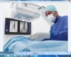
In that realm, Siemens holds a stake in the ViewRay system, an integrated MRI and Cobalt therapy system that lets physicians examine tumors in the body in real-time with MRI as they guide radiation therapy procedures. The system received FDA clearance in May.
MRI is also being integrated into the OR setting. Philips’ Ingenia MR-OR calls for an adjoining MRI chamber and OR that lets a doctor check mid-surgery if a patient’s tumor has been fully removed.
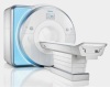
3T v. 1.5T
Channels and gradients on scanners can also be upgraded and customized depending on how advanced providers want to get with imaging. OEMs report that customers are starting to use the 3T scanners, which have greater signal strength and can capture more details, to image the liver, breast, pelvis and heart. But products, such as Philips’ MultiTransmit, are needed to improve images affected by dielectric effects, or shading on an image, which can occur when moving from a 1.5T up to a 3T.
According to Narayan, although more people are starting to look at 3T, 1.5T is still the clinical workhorse in a standard imaging center or hospital. “I don’t know if anytime in the near future it will surpass 1.5 or be equal. It’s a 60-40 split right now,” he says. Philips sees a similar split, but Mitchell says the trend shows a year over year increase for the number of 3T systems ordered.