by
Nancy Ryerson, Staff Writer | October 02, 2013
From the October 2013 issue of HealthCare Business News magazine
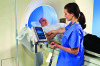
One recent study applied tract-based spatial statistics to perform a whole-brain diffusion tensor imaging (DTI) analysis of individuals at a clinical high risk of psychosis. DTI is a relatively new MRI technique that quantifies random water movement in the brain to determine if there is something abnormal in the connectivity of the brain’s white matter. The study found that the high-risk patients had an abnormal white matter microstructure.
“Schizophrenia is hypothesized to be a disorder of brain connectivity, which means that individual brain regions might function well individually, but do not properly work together as a team, like an orchestra without a conductor,” study author Christian Clemm von Hohenberg tells DOTmed Business News via email.


Ad Statistics
Times Displayed: 137680
Times Visited: 7952 MIT labs, experts in Multi-Vendor component level repair of: MRI Coils, RF amplifiers, Gradient Amplifiers Contrast Media Injectors. System repairs, sub-assembly repairs, component level repairs, refurbish/calibrate. info@mitlabsusa.com/+1 (305) 470-8013
Clemm von Hohenberg says MRI can help the treatment of diseases like schizophrenia become more biological, rather than be based on clinically observed symptoms. Besides DTI, he predicts more MRI techniques will be used for examining psychological issues.
“One very interesting challenge in the future is to combine more extensively different MRI techniques,” writes Clemm von Hohenberg. “These techniques (such as fMRI, DTI, MRI spectroscopy) provide different yet complementary information on brain structure and function. Combining this information will be crucial for understanding the human brain in health and disease.”
GE Healthcare has been involved in using that kind of technology to help protect one of the United States’ national resources: its football players. The company launched its $40 million “Head Health Initiative” earlier this year, with the goal of better understanding concussions and traumatic brain injury through imaging.
“Mild trajumatic brain injury is as very difficult thing to really kind of get on MR,” says GE’s Hausmann. “You need to pull all the technologies that you have, stability imaging to look at morphologic changes, metabolic changes, diffusion weighted imaging. It’s a subtle disease.”
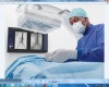
MR is also being used increasingly during brain surgery to help doctors adjust treatment midway through the procedure rather than after. The IMRIS operating suite, which includes MRI, has been used in 10,000 procedures since being released eight years ago.
“They can get feedback during the surgery,” says Jay D. Miller, president and CEO of IMRIS. “They can fix what they see as a potential problem, which is especially important for brain surgery. They want to make sure they get all of the tumor while the brain flap is still open.”